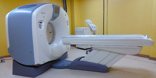
GE 128 slice CT scanner which paves the way for the future. The whole body scanner with a 0.35-second rotation speed which reduces kinetic artefact, examination time and contrast volume for vascular exploration dosage, while maintaining excellent diagnostic imagery.
The 750mm aperture maximises patients' comfort, reduces anxiety and stress, and enables effortless examinations.
Whole body scanning (1,700mm) can be done in 10 sec.
0.35sec, BP 0.83, 5mm slice width, 500mA (175mAs), only 6.5 sec from lung ? pelvis with 0.35sec/rot
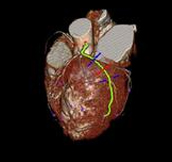
picture acquired together with electrocardiogram data. ECG triggering both retrospective and prospective mode possible.
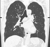
MPRs performed at oblique planes to the body or the coronary arteries.
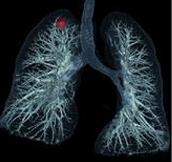
Lung area analysis software 3D display and automatic volume calculation for nodules.
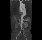
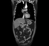
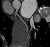
Body angiography with automatic bone and table removal - MIP reconstruction. Case: Aortic dissection. Left common iliac artery occlusion. Scanning: P0.83, 0.5s, 360mA, 100kV, weight 80kg, contrast 80cc
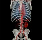
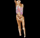
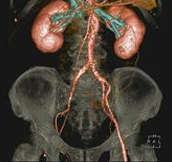
Body angiography with automatic table removal ? colored volume rendering reconstruction. Case: Aortic dissection. Left common iliac artery occlusion. Scanning: P0.83, 0.5s, 360mA, 100kV, weight 80kg, contrast 80cc
Save split image 2 x (512 x 512) matrix possible to see high resolution 3D image without doing partial reconstruction.

Save split image 2x(512 x 512) matrix possible to see high resolution 3D image without doing partial reconstruction.
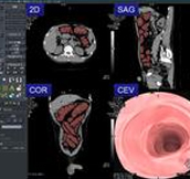
CT Colonoscopy with automatic colon area extraction from abdominal CT image. Display switchable to ?cruising?, ?3 section? and ? panorama mode.
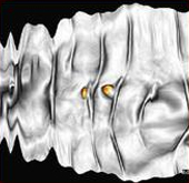
Color and display to process possible. Shape analysis filter (colored polyp on panoramic image)

0,5 sec/rotation, BP (BeamPitch) 1.08, 0.625mm slice width, 500mA (250mAs). High-pitch scanning possible for routine examination. 475 mm abdomen scannable within 5.5 seconds.
0.5 sec/rotation, BP 1.08, 0.625mm slice width, 360mA (180mAs).High-pitch scanning is possible for routine examination400 mm chest can be scanned within 4.7 seconds.

3D volume rendering reconstruction for separation of different soft tissues.

Tumor volume calculation in soft tissue.
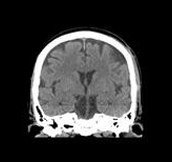
2.0 sec/rot, normal scanning, 0.625mm x 32i, 150mA. High image quality using normal scanning 64i mode for head MPR images


© 2016 Dr. Parvinder S. Lubana. All rights reserved | Developed & Design by Obabuji.com