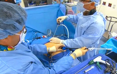
A laparoscopic cholecystectomy is a surgical procedure during which the doctor removes your gallbladder. This procedure uses several small incisions (cuts) instead of one large one.
A laparoscope, a narrow tube with a camera, is inserted through one incision. This allows your doctor to see your gallbladder on a TV screen. Your gallbladder is then removed through another small incision.

The procedure is used when you have stones in your gallbladder.
Your gallbladder stores bile, a fluid made by your liver. Bile helps digest fats in the foods you eat. Gallstones can block the flow of bile in your digestive system. This blockage can cause bloating, nausea, vomiting, and pain in your abdomen, shoulder, back, or chest. Gallstones can also block the ducts that channel the bile from the liver or gallbladder to the intestine. Gallstones can cause the gallbladder to become infected. A blockage in the common bile duct can cause jaundice (yellowing of your skin or eyes) or irritate the pancreas.
A general anesthetic is given to relax your muscles, prevent pain, and help you fall asleep. Your abdomen is inflated with carbon dioxide, a harmless gas. [A cholangiogram (a special X-ray) is done to check for stones in your common bile duct]. The laparoscope is then inserted through an incision in your navel, so your doctor can look inside.
Other instruments are then inserted through additional small incisions. Your gallbladder is removed through one of these incisions.
The risk of complications is very low, however, potential risks might include:
A cholecystectomy is the removal of your gallbladder through an incision in the upper abdomen.
An open cholecystectomy might be required instead of a laparoscopic cholecystectomy because of:
A general anesthetic is given to relax your muscles, prevent pain, and help you fall asleep. A single incision is made below the right side of your rib cage or in the center of the abdomen. Through the incision, your gallbladder and surrounding anatomy can be seen. The gallbladder is detached from its attachments, and the blood supply is tied off and divided. Sometimes a cholangiogram (a special X-ray) is conducted to check for stones in the common bile duct. If there are stones in the common bile duct, they are removed at this time. The skin is closed using surgical clips and sutures.
© Dr. Parvinder S. Lubana. All rights reserved. | Developed & Design by ipromptsolution.com